Spatial Biology Grants

Funding opportunities to support your next discovery

Apply to Win a Spatial Biology Grant

Submit your application by September 1, 2023

We’re dedicated to advancing research that grows our collective understanding of biology and contributes to the improvement of human health. If you have an interesting project that would benefit from spatial phenotyping, apply for a chance to win a spatial biology grant.

We are currently accepting applications for the 2023 Spatial Biology Discovery Grant, which is aimed at scientists pursuing immuno-oncology research.
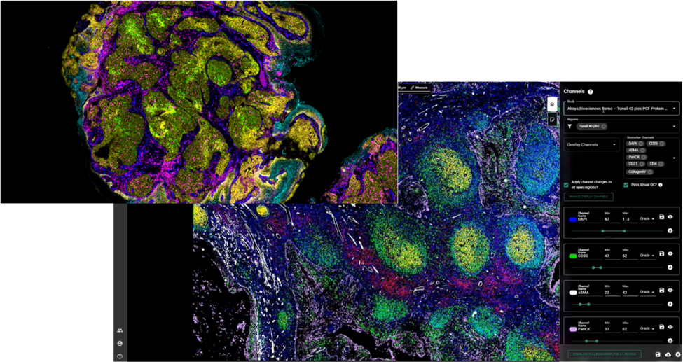
grant image
grant quote2

Providing opportunities for researchers involved in deep immune profiling projects to obtain a new perspective on their research through the power of PhenoCycler.

Frequently Asked Questions (FAQS)

A spatial biology grant is an opportunity for you to advance your research by obtaining rich spatial phenotyping data from your tissue samples for free. We host grant programs several times per year, and one or more awardees are selected for each program.

We partner with high-quality service providers to execute the staining, imaging for grant awardees. In most cases, all you have to do is send us your tissue sections, and we’ll send you back your spatial data!

Simply submit an application telling us about your project idea and how spatial phenotyping can help accelerate your research.

Grants open for submissions several times per year. The best way to stay informed is by bookmarking this page and checking back frequently.

Past Grants and Recipients

Fusion Spatial Multiomics Grant Program Winners

Howard-Crawford
Howard Crawford, P.h. D​
Scientific Director,​ Henry Ford Pancreatic Cancer Center​ Henry Ford Health
Daniel-Salas-Escabillas
Daniel Salas-Escabillas
5th year P.h.D Candidate, Cancer Biology University of Michigan / Henry Ford Pancreatic Cancer Center

100-plex Grant Program Winner

Ryan_Park_winner
Ryan Park, MD
Resident Physician
Harvard Radiation Oncology Program
Massachusetts General Hospital

Francis Crick Institute Spatial Biology Grant

Rute Ferreira, Postdoctoral Researcher, The Francis Crick Institute

Rute Ferreira
Postdoctoral Researcher
The Francis Crick Institute

Spatial Phenotyping Grant Program Winner

Ines-Sequira
Dr Inês Sequeira
Group Leader – Barts Charity Lecturer
Institute of Dentistry, Barts and The London School of Medicine and Dentistry
Queen Mary University of London

Spatial Multiomics Grant Award – PhenoCycler

BraunWinner
Dr. David Braun
Assistant Professor
Yale Center of Molecular and Cellular Oncology, Yale School of Medicine

Resources

Want to maximize your chances? We recommend you check out this spatial phenotyping whitepaper to help you outline in your abstract how a spatial approach will help accelerate your research.

At AACR 2023, we had the pleasure of catching up with Inês Sequeira, MSc, AFHEA, PhD from Queen Mary University of London – our first ever grant winner – and hearing from her about the awesome data she generated on the heterogeneity of oral cancers using her grant award. You can watch a recording of her poster presentation here.

CTA Header Copy Goes Here

Project Summary

The Akoya Biosciences Fusion Spatial Multiomics Grant Program was offered to researchers as an opportunity to explore the power of ultrahigh-plex deep spatial phenotyping with multi-analytes with early access to a groundbreaking new multiomics assay designed for the PhenoCycler-Fusion® platform. Our grant recipient was chosen based on their abstract describing the work they are performing at the Henry Ford Lab evaluating Patient Diversity and how that affects racial health disparities within pancreatic cancer patients.

Project Summary

Pancreatic ductal adenocarcinoma (PDAC) has exceptionally poor prognosis and is entirely resistant to current immunotherapies. PDAC has historically been split into 1) classical-like and 2) basal-like consensus subtypes, with the latter associated with worse survival. We recently refined this taxonomy (Hwang et al Nature Genetics in press) by developing a robust single-nucleus RNA-seq (snRNA-seq) approach to study a cohort of resected primary tumors and identified novel PDAC lineage subtypes associated with treatment-resistance and poor prognosis, including a “neural-like progenitor” (NRP) subtype that expresses genes normally found only in the brain. By using digital spatial profiling (NanoString GeoMx) on the resected tumors, we found that CD8 T cells are enriched in regions of tumor with NRP differentiation, which is surprising given that high CD8 T cell infiltration is generally associated with improved prognosis.

We propose to characterize the immune landscapes associated with each of the distinct PDAC malignant subtypes identified by our recent work. By applying the 100-plex Akoya panel to our resected primary PDAC tumors, we would identify the phenotype, cell state, and functionality of lymphoid and myeloid cells. We would use RNAscope on a consecutive section to classify the malignant cell subtypes based on transcription factor expression. In preliminary analysis of our GeoMx and snRNA-seq data, we have observed marked differences in the immune landscapes associated with each PDAC malignant subtype, including differences in expression levels of beta-2-microglobulin and HLA, cytokines, and genes associated with CD8 T cell effector function, differentiation, and exhaustion.

The Akoya platform would give us the opportunity to extend these spatial studies to the single-cell and protein level in order to deeply characterize the intra-tumoral heterogeneity in the tumor immune microenvironment of PDAC, revealing potentially distinct mechanisms of immune evasion that can inform combinatorial therapeutic strategies and thereby advance precision oncology for this lethal disease.

Project Summary

Congratulations to Rute Ferreira on the award of the Francis Crick Spatial Biology Grant. Rute will use this grant towards her goal in understanding how pancreatic cancer nerves regulate, and are regulated by, the cancer-associated inflammation using multispectral imaging with the PhenoImager HT in the Experimental Histopathology core unit.

Project Summary

To elucidate the functional and spatial intra-tumour heterogeneity of oral squamous cell carcinoma (OSCC) and to study the interaction of tumour cells with the microenvironment, in particular the role of specific mutations in promoting epithelial-to-mesenchymal transition and tumour progression.

Project Summary

My prior work utilized droplet-based scRNA-seq to understand how the immune microenvironment of kidney cancer (renal cell carcinoma) evolves with advancing disease stage (Braun et al, Cancer Cell, 2021). We identified a bi-directional “immune dysfunction circuit” of cell-cell interactions between CD8+ T cells and macrophages that was associated with a worse prognosis. This work highlighted the critical important of spatial context in understanding the tumor-immune microenvironment. We have now generated a robust scRNA-seq (and CITE-seq) dataset of the tumor-immune microenvironment for kidney cancer patients treated with immune checkpoint inhibitors, and have detailed response/resistance annotation for each. This work seeks to understand what features of the tumor-immune microenvironment impact response or resistance to current immunotherapy, with the hope that it will lead to novel approaches to combination immune-based therapeutic approaches. This grant award would allow simultaneous spatial characterization of already obtained pre-treatment tumor tissue, and would allow us to integrate with our droplet-based scRNA-seq dataset to understand how cell-cell interactions and cellular “neighborhoods” in the microenvironment contribute to response and resistance.