We had a great time in National Harbor, MD earlier this month for the 34th Annual Meeting of The Society for Immunotherapy of Cancer (SITC). We were excited to host a dinner symposium at the meeting, where we took the opportunity to share some exciting product updates and new data from our multiplexed immunofluorescence platforms. We heard from one of our customers, Dr. Robert Pierce from the Fred Hutchinson Cancer Research Center, who spoke about how his team uses Akoya’s platforms to study the tumor microenvironment (TME). There were also multiple posters on display with new data from our platforms.
If you couldn’t attend SITC this year, check out our recap of the event below.
Symposium Highlights
Our CEO, Brian McKelligon, started off the presentations with a quick introduction about Akoya, our history, and how the CODEX® and Phenoptics™ platforms complement each other to support research from discovery to the translational and clinical phases. CODEX® integrates with existing fluorescence microscopes to image up to 40 markers per tissue sample for hypothesis-free biomarker discovery. The Phenoptics™ platform is the industry standard for rapid, high throughput multiplexed IHC (mIHC).
We then heard from Victoria Duckworth, Product Manager for Phenoptics™ Panels and Kits, who introduced our new Opal™ MOTiF™ PD-1/PD-L1 Panel Kits. These six-plex, seven-color panels have been validated on melanoma and lung cancer samples, with ready-to-use primary and secondary antibodies automated for third-party autostainers. Victoria explained how same-day image analysis is made possible with pre-optimized acquisition protocols for the Vectra® Polaris™ System and pre-configured algorithms for the inForm® software package. Together, this workflow provides the only complete and turn-key multiplex IHC solution for immuno-oncology translational research.

If you’re interested in learning more, we’re hosting a webinar on Dec 17th to discuss our new panel kits and share some tips for customizing with your own antibodies. Register here.
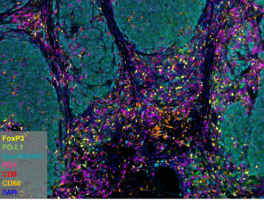
Opal™ MoTiF™ PD-1/PD-L1 Melanoma Panel
Next up was Cliff Hoyt, VP of Translational Programs and Scientific Affairs. His talk focused on cross-validation of biomarkers between CODEX® and Phenoptics™, so our customers can easily translate their findings across platforms as their studies move along the continuum from discovery to translational and clinical research.
Cliff revealed the results of a comparative study between CODEX® and Opal™ staining on serial sections of tonsil and lung cancer tissue samples. The 28-plex CODEX® panel included all markers from the 6-plex Opal™ panel. Both methods detected the same cell phenotypes and displayed similar marker frequencies, demonstrating that these two approaches can be made analytically equivalent. There were some slight differences in the results. For example, while PD-L1 was amplified in both methods and delivered equivalent results, PD-1 was amplified on Opal™, but not CODEX® , accounting for the difference in signal intensity. To address these discrepancies, Cliff described how we’ve added amplification to CODEX® detection through a novel amplification method.

This cross-platform comparison provides a conceptual framework for biomarkers discovered on the CODEX® platform to be translated to the Phenoptics™ platform for high-throughput translational studies. We will be launching a new CODEX® reagent offering in 2020 to amplify key immuno-oncology markers in the tumor microenvironment.
Our final speaker was Dr. Robert Pierce, the Scientific Director of the Immunopathology Lab at Fred Hutchinson Cancer Research Center. In his presentation, he described how he and his collaborators utilized the Phenoptics™ platform to glean important spatial insights into the tumor microenvironment. The focus of his talk was the immunosuppressive role of myeloid cells in the tumor microenvironment and in regulating treatment response.
His data showed that tumor-associated macrophages, or “badophages”, can distinguish prognosis in multiple tumor types and facilitate metastasis in the MISTRG melanoma model, a mouse model repopulated with a human immune system. In another study in non-small cell lung cancer (NSCLC), spatial analysis was used to identify a link between the ratio of CD8 to polymorphonuclear neutrophils (PMN) and treatment response to PD-1/PD-L1 inhibitors. Finally, Dr. Pierce also emphasized the utility of integrating single-cell RNA sequencing with multiplexed IHC to better understand the potential mechanisms of immunosubversion in the tumor microenvironment.
Our sincere thanks to everyone who attended our workshop. We hope to see you again next year!
Poster Synopses
P64 – HIGHLY MULTIPLEXED SINGLE-CELL SPATIAL ANALYSIS OF FFPE TUMOR TISSUES USING CODEX®
In this poster, 28-plex analysis of human FFPE tissues was performed with CODEX® to understand spatial interactions in the tumor microenvironment. The CODEX® workflow was adjusted to incorporate TSA-mediated dyes for the amplification of low-expressing markers, including FOXP3, PD-L1, and PD-1. Antibodies for these markers were run and imaged with both the standard CODEX® workflow and the new amplification workflow. Signal intensity for the amplified markers was significantly increased when compared to the standard CODEX® run. We plan to integrate these TSA-mediated dyes into the standard CODEX® workflow to enable researchers to study key low-expressing markers.

P34 – A FULLY OPTIMIZED END-TO-END SOLUTION FOR I/O MULTIPLEX IMMUNOFLUORESCENCE STAINING USING OPAL POLARIS 7-COLOR PD-1/PD-L1 PANEL KITS FOR LUNG CANCER AND MELANOMA
Our new MOTiF™ PD-1/PD-L1 Panel Kits were developed to address the need for a reproducible, simple, and standardized multiplex immunofluorescent assay that can assess spatial relationships between biomarkers. In this study, our automated MOTiF™ workflow was validated on lung cancer and melanoma FFPE samples. The kits integrate with a pre-configured whole slide multispectral imaging and analysis workflow (on the Vectra® Polaris™ and InForm® software) to provide the only validated end-to-end solution for quantitative data for translational immuno-oncology research.

P43 – A NOVEL PLATFORM FOR HIGHLY MULTIPLEXED, SINGLE-CELL IMAGING OF CELL SUSPENSIONS
This poster, a collaboration between Akoya and researchers at Stanford University, was a preliminary study into the compatibility of the CODEX® system with cell suspensions. An in-depth immune profile of PBMC samples was generated using CODEX® , with PBMCs fixed and spread on a coverslip and stained using 26 different markers. CODEX® performed well, with most markers displaying good signal/noise separation. Staining patterns were qualitatively assessed: mutually exclusive markers showed no overlap in staining, and co-expression was observed in similar cell types. Signal intensity also remained relatively constant over 9 cycles. With validation and optimization of markers for cell suspensions, staining performance could improve further. The results from this initial study indicate that CODEX® can be used as a faster, simpler, and more affordable alternative for cellular biomarker analysis.

P31 – APPLYING MULTISPECTRAL UNMIXING AND SPATIAL ANALYSES TO EXPLORE TUMOR HETEROGENEITY WITH A PRE-OPTIMIZED 7-COLOR IMMUNO-ONCOLOGY WORKFLOW
We characterized the tumor heterogeneity of lung cancer samples using our new end-to-end translational workflow based on the Phenoptics™ platform. The MOTiF™ PD-1/PD-L1 Panel Kits include six clinically relevant antibodies and their Opal fluorophores. In combination with pre-defined parameters for multispectral scanning on the Vectra® Polaris™ and a pre-configured algorithm for the inForm® software, MOTiF™ provides an end-to-end solution from staining to analysis. The heterogeneity of spatial interactions was demonstrated among different lung cancer samples using tissue microarrays (TMA). Multispectral imaging (on Vectra® Polaris™) was shown to improve sensitivity of detection through spectral unmixing, and the full workflow from staining, imaging, unmixing, and spatial analysis provides a robust platform for analysis of the tumor microenvironment.


