Main Menu
We’ve rebranded some of our products, learn more ›
CODEX® is now PhenoCycler,
Phenoptics™ is now Phenolmager.
Validating Multiplexed Antibody Panels for Pancreatic Research Applications
Recorded on Nov 7, 2019
Validating CODEX Panels for Pancreatic Research
Studying the pathogenesis of diabetes requires detailed analysis of the pancreatic islet microenvironment and its numerous cell types, at the single-cell level. Profiling these complex cellular phenotypes is greatly enhanced by the application of highly multiplexed immunofluorescence technology that can also retain the spatial context of the entire tissue section.
The CODEX® System is a compact, budget-friendly instrument that integrates with third-party fluorescence microscopes and images 40+ protein markers in a single tissue section. The platform is part of an end-to-end solution that also includes inventoried antibodies, reagents, and a software solution for visualizing cellular phenotypes. In the Powers and Brissova Research Group at Vanderbilt University, the CODEX platform is used to investigate unique cellular niches in the developing human pancreas, including describing the interactions between endocrine cells and neurovascular and immune compartments.
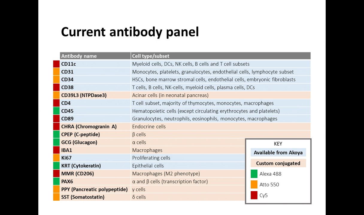
In this webinar, Diane Saunders, a staff scientist in the Vanderbilt Diabetes Center, will describe the validation process for custom panels of CODEX antibodies using lightly fixed tissue samples. She will also share the preliminary results from her validation studies, where she used a multiplexed CODEX panel with a mix of preconjugated and custom-conjugated antibodies, to profile pancreatic islet development in neonatal human samples.
Grady Carlson, a Technical Application Scientist at Akoya Biosciences, will introduce the CODEX technology and workflow and how it can be applied for biomarker discovery.
Learning objectives:
- Outlining tips & tricks to validate highly multiplexed antibody panels for lightly-fixed pancreatic tissue
- Demonstrating why spatially-resolved, multiplexed immunofluorescence is essential for tissue analysis and biomarker discovery
- Investigating how the CODEX solution enables the simultaneous analysis of 40+ proteins in FFPE and fresh frozen tissue sections
Video

Speakers

Dr. Diane Saunders
Staff Scientist, Powers & Brissova Research Group Vanderbilt University Medical Center

Dr. Grady Carlson
Technical Application Scientist Akoya Biosciences


