

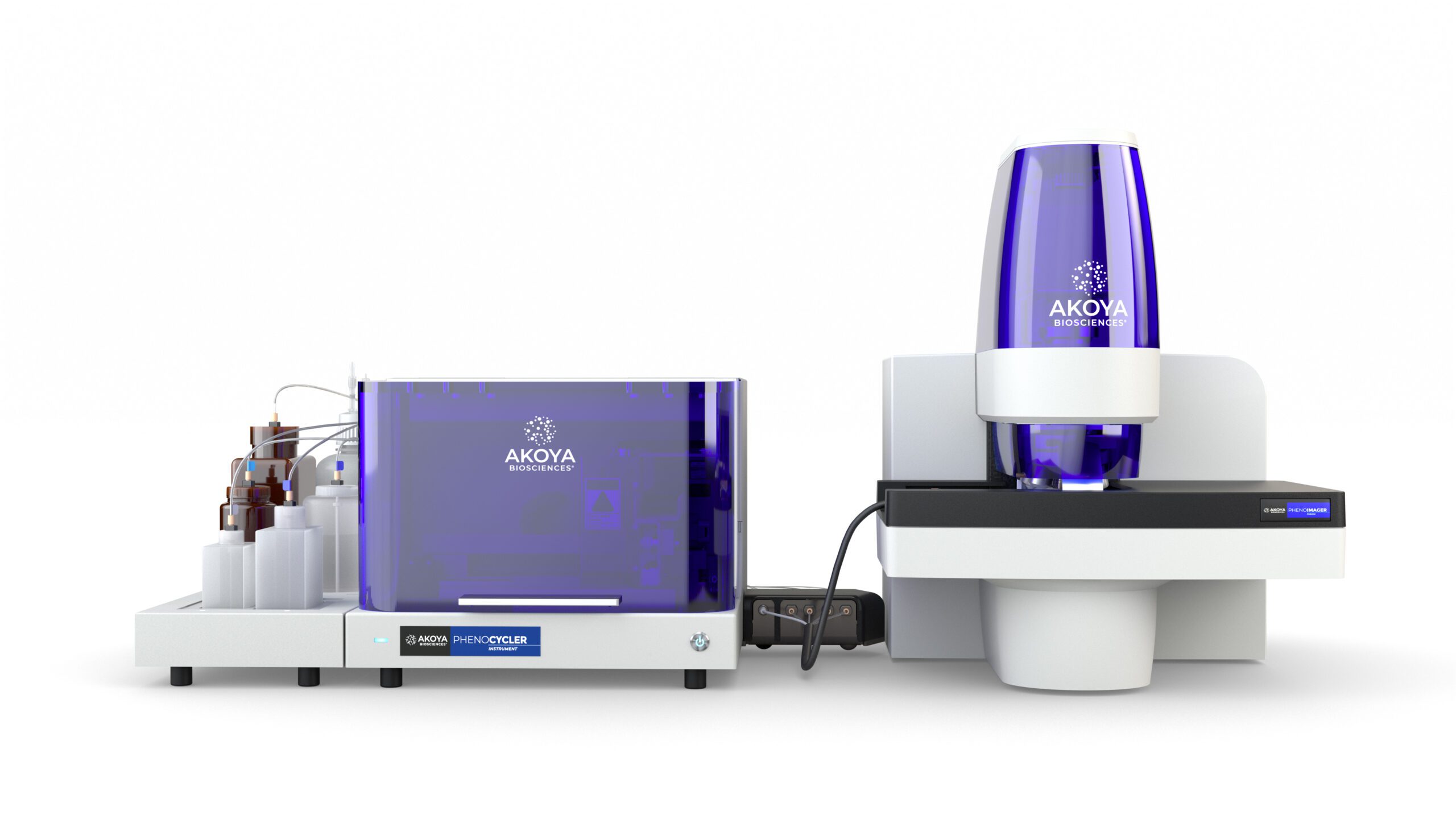
The PhenoCycler-Fusion 2.0 Solution
Spatial Discoveries at YOUR Scale
We’ve rebranded some of our products, learn more ›
CODEX® is now PhenoCycler,
Phenoptics™ is now Phenolmager.
Experience the Fastest Spatial Biology Solution
Whether you're unlocking insights from a single sample, conducting spatial studies on dozens of samples, or exploring population-level analyses on hundreds of samples, PhenoCycler®-Fusion 2.0 will match the scale of your spatial studies.
Powered by high-speed imaging and proprietary barcoding technology, PhenoCycler-Fusion 2.0 now introduces multi-slide automation with parallel processing of slides. This advanced capability amplifies your ability to uncover more discoveries faster than ever before.
Why choose PhenoCycler-Fusion 2.0?
Faster than Ever
Image 1 million cells in 10 minutes with accelerated parallel imaging and fluidics. Experience a new standard for speed and time to results faster than ever before.
High-throughput Excellence
Maximize your weekly throughput without compromising imaging area, plex, or data. Effortlessly phenotype 40 samples at 20-plex or 300 samples at 6-plex.
Scalable Plexing
Unlock scalable plexing with our barcoded antibody technology. This innovative approach combines antibody specificity with molecular barcodes enabling simultaneous detection of over 100 targets at high spatial resolution while preserving tissue integrity.
Unmatched Content
Design your desired panels from a database of 350+ antibodies or save time with ready-to-use PhenoCode™ Discovery Panels. Explore beyond the limitations of species and epitope degradation.
Explore the Data Firsthand
Hear from Our Users


PhenoCycler-Fusion 2.0 Technology
PhenoCycler-Fusion 2.0 capitalizes on our innovation and expertise in high-speed imaging and automated cycling, emerging as the fastest spatial biology solution with unparalleled throughput.

A Simple End-to-End Workflow
Stain

Image

Analyze

Integrate

Seminal Publications
Single-cell Spatial Metabolic and Immune Phenotyping of Head and Neck Cancer Tissues Identifies Tissue Signatures of Response and Resistance to Immunotherapy
A Spatially Resolved Single-cell Genomic Atlas of the Adult Human Breast
Coordinated Cellular Neighborhoods Orchestrate Antitumoral Immunity at the Colorectal Cancer Invasive Front
Explore More

