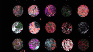
We recently held a social media image contest inviting our followers to show off a fun activity or theme using tissue images. After receiving some incredibly creative submissions, it’s time to announce the winners! Here are the top three participants with the most likes and/or reactions on their social media posts.
1st Place: Conrad Lee, The Chinese University of Hong Kong
Conrad Lee, from the Chinese University of Hong Kong, shared this beautiful image of FFPE breast tissue which was stained for cytokeratin and CD8 and imaged using the Phenoptics™ platform. The pattern of red and green stains looks just like blooming red roses.

2nd Place: Ayse Akarca, University College London
Ayse Akarca is a research fellow at the University College London. She captured this image of kidney tissue using the Phenoptics platform and found that it’s shaped just like a turtle!

3rd Place: Claire Alexandra, Manchester Cancer Research Centre
Claire Alexandra, from the Manchester Cancer Research Centre, took this colorful image of human prostate tissue (left) using the Phenoptics platform. It looks very similar to another image she captured: this impressive astronomical photo of the surface of the sun (right).

Congratulations to all the winners! We loved seeing all the submissions that came in – you can check them out too by scrolling through the #AkoyaImageContest tag on Twitter and LinkedIn.
Want to take your own multiplex immunofluorescence images? Learn about our spatial biology solutions.