A Launch at SITC 2022
Akoya recently wrapped up two very exciting back-to-back conferences. Each conference catered to specific audiences, and each offered a unique Akoya experience.
The first conference in chronological order was the Society for Immunotherapy of Cancer (SITC) meeting, which was held in Boston Massachusetts this year. SITC can be considered a relatively large conference taking place November 8th to the 12th, it typically brings together over 5,000 people including research scientists, physician scientists, clinicians, patients, patient advocates, government representatives and industry leaders. The goal of this conference is to foster scientific exchange and collaboration; focus on initiatives of major importance in the field and overall bring together all aspects of the cancer immunology and immunotherapy community. Currently, SITC has more than 4,650 members who represent over 35 medical specialties in 63 countries around the world.
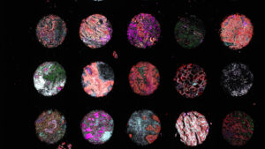
What we at Akoya were most excited about was taking the opportunity that this conference offers to launch our PhenoCode™ Signature Panels. Briefly, immune checkpoints are established therapeutic targets for cancer treatment, and it is increasingly thought that combination therapies, which target multiple checkpoints at once, lead to improved patient outcomes. The development of clinically relevant prognostic tools to stratify patients will be critical for the advancement of such treatments. Spatial Phenotyping of the tumor microenvironment (TME) by assessing both, protein co-expression and spatial relationships between cells, appears to improve patient stratification. To provide researchers with an automated end-to-end workflow for the functional evaluation of tumors and stratification of patients, we have thus developed a new single-cell multiplexed staining solution. PhenoCode signature panels are readymade antibody panels that enable comprehensive analysis of the tumor microenvironment. They are designed to provide flexibility, allowing for the easy integration of one additional marker to a given 5-plex panel. Some panels include an immune profile panel, the activated tumor infiltrating lymphocyte panel, the immune-contexture panel, the M1/M2 polarization panel, and the exhaustion panel. Each panel is optimized and validated to enable the rapid development of spatial signatures by shortening the assay development times by three times giving user more time analyzing data and less time preparing the experiment.

Our proud Akoya team gathered at the SITC booth. The poster presented at SITC can be downloaded here
Each of the customizable multiplex panels includes key markers for phenotyping the TME and immune status. When combined with the high-speed and robust imaging of the PhenoImager platforms, a rapid, quantitative, end-to-end spatial phenotyping workflow is enabled. The workflow accelerates development and validation of predictive signatures and prognostic biomarkers for immuno-oncology applications. The PhenoCode Signature chemistry represents an important milestone in Akoya’s commitment to create a suite of platform solutions from discovery to diagnostics. As a powerful hybrid of Akoya’s legacy CODEX and Opal assays, the PhenoCode Signature Panels create a continuum of methodologies, enabling translational and clinical researchers to perform discovery on the PhenoCycler®-Fusion and validation on the PhenoImager platforms.
During the conference, aside from one-on-one conversations within the booth we also delivered several presentations and a symposium describing the application of PhenoCode Signature Panels given by Patrick Savickas PhD, Immunochemist, HistoWiz, Arutha Kulasinghe, PhD, Senior Research Fellow, University of Queensland, and Oliver Braubach, PhD, Head of Applications, Akoya Biosciences. To get some of the details presented during this conference please feel free to attend our virtual launch event. Register here
Some Exciting Neuro Data at SFN 2022
The next conference was the Society for Neuroscience conference (SFN) which took place on the opposite side of the country in San Diego California from November 12th to the 16th. Oliver Braubach is our Director of Applications at Akoya and a classically trained neuroscientist, so with this content having a special place in his heart (or should we say his brain) the focus was all about data, data, and more neuro data. The work the applications team focused on was investigating the microglia in human brain tissue derived from patients with Alzheimer’s disease. Neurological diseases remain a developing arm of the spatial biology realm but the need to research these conditions is nevertheless extremely important and urgent. Neurological diseases ranging from neurodegenerative diseases to cancer often have poor prognoses and are associated with a high cost of care. According to the Centers for Disease Control and Prevention (CDC) Alzheimer’s disease is the most common form of dementia, the 6th leading cause of death among adults in the United States, and among the top 10 overall leading causes of death. Neuroinflammation is increasingly linked to the etiology and progression of Alzheimer’s disease (AD). Microglia, the critical stewards of the brain’s immune microenvironment, display phenotypic and state diversity in AD as shown in single-cell transcriptomic and proteomic analyses on tissue homogenates. These approaches lack spatial information, which is crucial to interpreting microglial involvement in neuroinflammation.

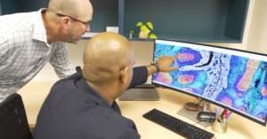
Standing by at SFN to give presentation on neuro data is Harry Lui, Senior Research Associate and first author of our poster. The tissue used to generate these data can be viewed in our data gallery and the poster can be viewed here
For this conference we showed our data in a poster presentation that focused on the development of a workflow that provides both, deep biomarker readouts and excellent spatial resolution. To this end, we developed a PhenoCycler®–Fusion (PCF) workflow that is compatible with formalin-fixed human brain tissue. The PCF is a novel spatial biology platform that generates ultrahigh-plex spatial data for millions of cells on a single tissue section within a matter of hours. Using this technology, we characterized the immune microenvironment – with emphasis on microglia – in normal and AD brain tissues. These findings suggest an enrichment of certain microglial subtypes in AD brain tissues and in proximity to Ab plaques. These exciting data provide a method for ultrahigh-plex immunofluorescent analyses of AD pathology and new insights into the inflammatory microenvironment of AD.
Author: James DeRosa, MPH